

What Are The Types Of Tissues In Animals
The Animal Body: Bones Class and Function
174 Animal Master Tissues
Learning Objectives
By the terminate of this section, you will be able to practice the following:
- Describe epithelial tissues
- Hash out the dissimilar types of connective tissues in animals
- Describe three types of musculus tissues
- Describe nervous tissue
The tissues of multicellular, complex animals are four primary types: epithelial, connective, muscle, and nervous. Think that tissues are groups of similar cells (cells conveying out related functions). These tissues combine to form organs—like the skin or kidney—that have specific, specialized functions inside the trunk. Organs are organized into organ systems to perform functions; examples include the circulatory system, which consists of the centre and blood vessels, and the digestive system, consisting of several organs, including the stomach, intestines, liver, and pancreas. Organ systems come together to create an unabridged organism.
Epithelial Tissues
Epithelial tissues cover the outside of organs and structures in the body and line the lumens of organs in a single layer or multiple layers of cells. The types of epithelia are classified by the shapes of cells present and the number of layers of cells. Epithelia composed of a single layer of cells is chosen unproblematic epithelia; epithelial tissue composed of multiple layers is chosen stratified epithelia. (Figure) summarizes the different types of epithelial tissues.
| Different Types of Epithelial Tissues | ||
|---|---|---|
| Cell shape | Description | Location |
| squamous | apartment, irregular round shape | unproblematic: lung alveoli, capillaries; stratified: skin, mouth, vagina |
| cuboidal | cube shaped, cardinal nucleus | glands, renal tubules |
| columnar | tall, narrow, nucleus toward base; tall, narrow, nucleus along cell | simple: digestive tract; pseudostratified: respiratory tract |
| transitional | circular, simple but appear stratified | urinary bladder |
Squamous Epithelia
Squamous epithelial cells are mostly round, flat, and have a small, centrally located nucleus. The cell outline is slightly irregular, and cells fit together to form a covering or lining. When the cells are arranged in a single layer (simple epithelia), they facilitate diffusion in tissues, such as the areas of gas exchange in the lungs and the exchange of nutrients and waste matter at claret capillaries.
Squamous epithelia cells (a) have a slightly irregular shape, and a small, centrally located nucleus. These cells tin can be stratified into layers, equally in (b) this human cervix specimen. (credit b: modification of piece of work by Ed Uthman; scale-bar data from Matt Russell)
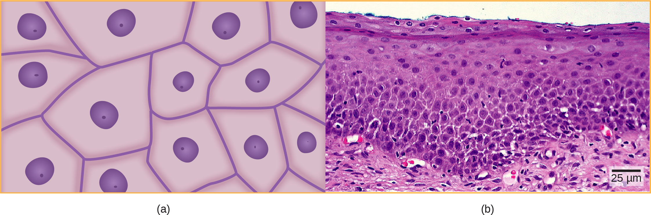
(Figure)a illustrates a layer of squamous cells with their membranes joined together to class an epithelium. Image (Figure)b illustrates squamous epithelial cells arranged in stratified layers, where protection is needed on the body from exterior abrasion and harm. This is chosen a stratified squamous epithelium and occurs in the pare and in tissues lining the mouth and vagina.
Cuboidal Epithelia
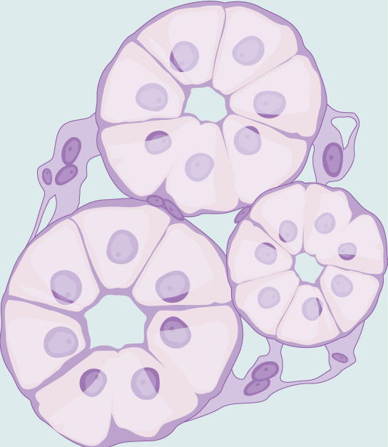
Cuboidal epithelial cells, shown in (Figure), are cube-shaped with a single, central nucleus. They are well-nigh normally constitute in a single layer representing a elementary epithelia in glandular tissues throughout the body where they set up and secrete glandular material. They are also found in the walls of tubules and in the ducts of the kidney and liver.
Unproblematic cuboidal epithelial cells line tubules in the mammalian kidney, where they are involved in filtering the blood.
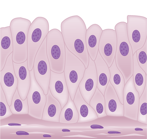
Columnar Epithelia
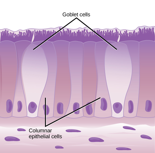
Columnar epithelial cells are taller than they are wide: they resemble a stack of columns in an epithelial layer, and are most commonly found in a single-layer arrangement. The nuclei of columnar epithelial cells in the digestive tract announced to be lined upward at the base of the cells, every bit illustrated in (Effigy). These cells blot material from the lumen of the digestive tract and prepare it for entry into the body through the circulatory and lymphatic systems.
Simple columnar epithelial cells absorb textile from the digestive tract. Goblet cells secrete mucous into the digestive tract lumen.
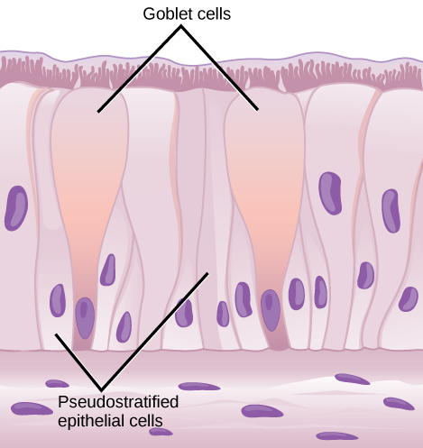
Columnar epithelial cells lining the respiratory tract appear to be stratified. However, each cell is fastened to the base membrane of the tissue and, therefore, they are simple tissues. The nuclei are arranged at unlike levels in the layer of cells, making it appear equally though there is more than one layer, as seen in (Figure). This is called pseudostratified, columnar epithelia. This cellular covering has cilia at the upmost, or free, surface of the cells. The cilia enhance the movement of mucous and trapped particles out of the respiratory tract, helping to protect the system from invasive microorganisms and harmful material that has been breathed into the body. Goblet cells are interspersed in some tissues (such every bit the lining of the trachea). The goblet cells comprise mucous that traps irritants, which in the instance of the trachea keep these irritants from getting into the lungs.
Pseudostratified columnar epithelia line the respiratory tract. They exist in 1 layer, but the arrangement of nuclei at unlike levels makes it appear that there is more i layer. Goblet cells interspersed between the columnar epithelial cells secrete mucous into the respiratory tract.
Transitional Epithelia
Transitional or uroepithelial cells announced simply in the urinary arrangement, primarily in the bladder and ureter. These cells are arranged in a stratified layer, but they have the capability of appearing to pile up on pinnacle of each other in a relaxed, empty bladder, equally illustrated in (Figure). Every bit the urinary float fills, the epithelial layer unfolds and expands to concur the volume of urine introduced into information technology. Equally the float fills, it expands and the lining becomes thinner. In other words, the tissue transitions from thick to thin.
Visual Connectedness
Transitional epithelia of the urinary bladder undergo changes in thickness depending on how full the bladder is.
Which of the following statements about types of epithelial cells is simulated?
- Simple columnar epithelial cells line the tissue of the lung.
- Simple cuboidal epithelial cells are involved in the filtering of blood in the kidney.
- Pseudostratisfied columnar epithilia occur in a single layer, but the arrangement of nuclei makes it appear that more than one layer is present.
- Transitional epithelia alter in thickness depending on how full the bladder is.
<!–<para>A–>
Connective Tissues
Connective tissues are fabricated upwardly of a matrix consisting of living cells and a nonliving substance, called the ground substance. The ground substance is fabricated of an organic substance (usually a protein) and an inorganic substance (usually a mineral or water). The principal cell of connective tissues is the fibroblast. This cell makes the fibers institute in nearly all of the connective tissues. Fibroblasts are motile, able to carry out mitosis, and can synthesize whichever connective tissue is needed. Macrophages, lymphocytes, and, occasionally, leukocytes can be found in some of the tissues. Some tissues have specialized cells that are not found in the others. The matrix in connective tissues gives the tissue its density. When a connective tissue has a high concentration of cells or fibers, it has proportionally a less dense matrix.
The organic portion or protein fibers found in connective tissues are either collagen, elastic, or reticular fibers. Collagen fibers provide strength to the tissue, preventing it from being torn or separated from the surrounding tissues. Elastic fibers are fabricated of the protein elastin; this fiber tin stretch to one and one half of its length and render to its original size and shape. Elastic fibers provide flexibility to the tissues. Reticular fibers are the third type of poly peptide fiber institute in connective tissues. This cobweb consists of thin strands of collagen that form a network of fibers to back up the tissue and other organs to which it is continued. The various types of connective tissues, the types of cells and fibers they are fabricated of, and sample locations of the tissues is summarized in (Figure).
| Connective Tissues | |||
|---|---|---|---|
| Tissue | Cells | Fibers | Location |
| loose/areolar | fibroblasts, macrophages, some lymphocytes, some neutrophils | few: collagen, elastic, reticular | effectually blood vessels; anchors epithelia |
| dense, fibrous connective tissue | fibroblasts, macrophages | mostly collagen | irregular: skin; regular: tendons, ligaments |
| cartilage | chondrocytes, chondroblasts | hyaline: few: collagen fibrocartilage: large amount of collagen | shark skeleton, fetal bones, human ears, intervertebral discs |
| bone | osteoblasts, osteocytes, osteoclasts | some: collagen, rubberband | vertebrate skeletons |
| adipose | adipocytes | few | adipose (fat) |
| claret | reddish blood cells, white blood cells | none | blood |
Loose/Areolar Connective Tissue
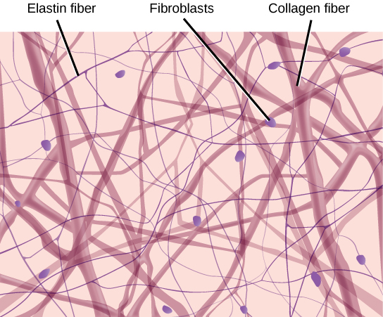
Loose connective tissue, also called areolar connective tissue, has a sampling of all of the components of a connective tissue. As illustrated in (Figure), loose connective tissue has some fibroblasts; macrophages are present also. Collagen fibers are relatively wide and stain a light pink, while elastic fibers are thin and stain night blueish to black. The infinite between the formed elements of the tissue is filled with the matrix. The cloth in the connective tissue gives it a loose consistency similar to a cotton ball that has been pulled apart. Loose connective tissue is establish around every blood vessel and helps to proceed the vessel in place. The tissue is also constitute around and betwixt most body organs. In summary, areolar tissue is tough, withal flexible, and comprises membranes.
Loose connective tissue is composed of loosely woven collagen and elastic fibers. The fibers and other components of the connective tissue matrix are secreted past fibroblasts.
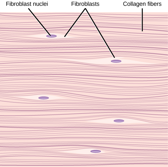
Gristly Connective Tissue
Fibrous connective tissues incorporate large amounts of collagen fibers and few cells or matrix material. The fibers tin can be arranged irregularly or regularly with the strands lined up in parallel. Irregularly arranged fibrous connective tissues are constitute in areas of the torso where stress occurs from all directions, such every bit the dermis of the skin. Regular fibrous connective tissue, shown in (Figure), is found in tendons (which connect muscles to basic) and ligaments (which connect bones to bones).
Fibrous connective tissue from the tendon has strands of collagen fibers lined up in parallel.
Cartilage
Cartilage is a connective tissue with a big corporeality of the matrix and variable amounts of fibers. The cells, called chondrocytes, make the matrix and fibers of the tissue. Chondrocytes are found in spaces within the tissue called lacunae.
A cartilage with few collagen and elastic fibers is hyaline cartilage, illustrated in (Effigy). The lacunae are randomly scattered throughout the tissue and the matrix takes on a milky or scrubbed appearance with routine histological stains. Sharks have cartilaginous skeletons, every bit does nearly the entire human skeleton during a specific pre-birth developmental phase. A remnant of this cartilage persists in the outer portion of the human nose. Hyaline cartilage is likewise constitute at the ends of long basic, reducing friction and cushioning the articulations of these bones.
Hyaline cartilage consists of a matrix with cells called chondrocytes embedded in it. The chondrocytes be in cavities in the matrix chosen lacunae.
Elastic cartilage has a large corporeality of elastic fibers, giving it tremendous flexibility. The ears of near vertebrate animals contain this cartilage as do portions of the larynx, or vox box. Fibrocartilage contains a large amount of collagen fibers, giving the tissue tremendous strength. Fibrocartilage comprises the intervertebral discs in vertebrate animals. Hyaline cartilage found in movable joints such every bit the knee joint and shoulder becomes damaged as a result of age or trauma. Damaged hyaline cartilage is replaced by fibrocartilage and results in the joints condign "stiff."
Bone
Bone, or osseous tissue, is a connective tissue that has a large amount of two different types of matrix cloth. The organic matrix is similar to the matrix material found in other connective tissues, including some amount of collagen and elastic fibers. This gives strength and flexibility to the tissue. The inorganic matrix consists of mineral salts—by and large calcium salts—that give the tissue hardness. Without acceptable organic fabric in the matrix, the tissue breaks; without acceptable inorganic material in the matrix, the tissue bends.
In that location are three types of cells in bone: osteoblasts, osteocytes, and osteoclasts. Osteoblasts are active in making bone for growth and remodeling. Osteoblasts deposit os material into the matrix and, after the matrix surrounds them, they go along to alive, only in a reduced metabolic land as osteocytes. Osteocytes are establish in lacunae of the bone. Osteoclasts are agile in breaking downwards bone for bone remodeling, and they provide access to calcium stored in tissues. Osteoclasts are usually plant on the surface of the tissue.
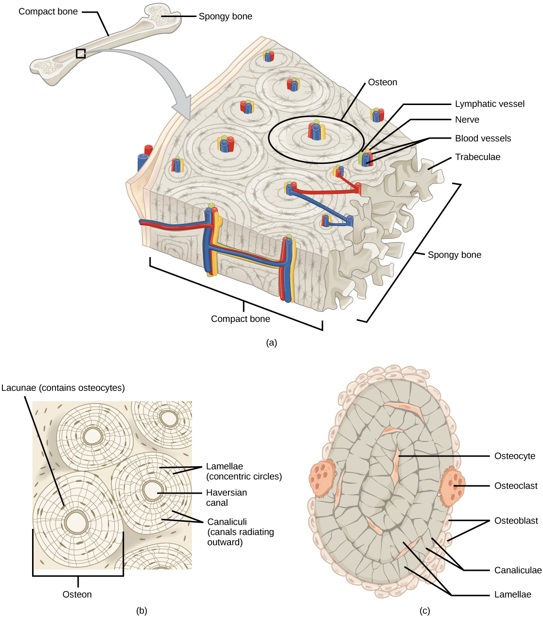
Bone can exist divided into two types: compact and spongy. Compact bone is found in the shaft (or diaphysis) of a long bone and the surface of the flat bones, while spongy bone is constitute in the end (or epiphysis) of a long bone. Compact bone is organized into subunits called osteons, equally illustrated in (Figure). A blood vessel and a nerve are establish in the center of the construction within the Haversian canal, with radiating circles of lacunae around information technology known as lamellae. The wavy lines seen between the lacunae are microchannels called canaliculi; they connect the lacunae to aid improvidence between the cells. Spongy bone is fabricated of tiny plates chosen trabeculae; these plates serve as struts to give the spongy bone strength. Over time, these plates can intermission causing the os to become less resilient. Bone tissue forms the internal skeleton of vertebrate animals, providing structure to the brute and points of attachment for tendons.
(a) Meaty os is a dense matrix on the outer surface of bone. Spongy bone, inside the compact bone, is porous with spider web-like trabeculae. (b) Meaty bone is organized into rings called osteons. Blood vessels, nerves, and lymphatic vessels are institute in the fundamental Haversian canal. Rings of lamellae surround the Haversian canal. Between the lamellae are cavities called lacunae. Canaliculi are microchannels connecting the lacunae together. (c) Osteoblasts surround the exterior of the os. Osteoclasts bore tunnels into the bone and osteocytes are establish in the lacunae.
Adipose Tissue
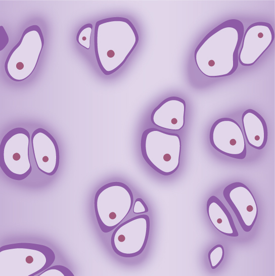
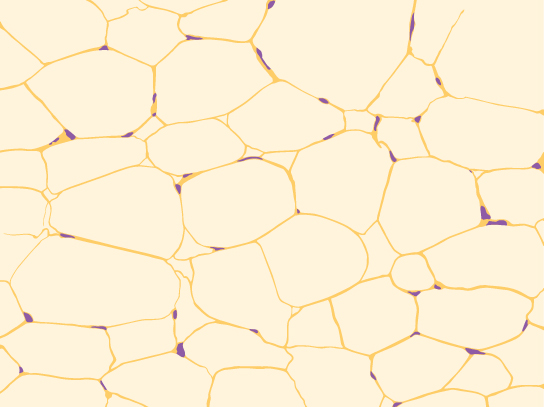
Adipose tissue, or fat tissue, is considered a connective tissue even though it does not take fibroblasts or a real matrix and but has a few fibers. Adipose tissue is made upwards of cells called adipocytes that collect and store fat in the course of triglycerides, for energy metabolism. Adipose tissues additionally serve as insulation to help maintain body temperatures, allowing animals to be endothermic, and they function as cushioning against impairment to body organs. Under a microscope, adipose tissue cells appear empty due to the extraction of fat during the processing of the textile for viewing, as seen in (Figure). The thin lines in the image are the cell membranes, and the nuclei are the small, black dots at the edges of the cells.
Adipose is a connective tissue is made up of cells chosen adipocytes. Adipocytes have pocket-size nuclei localized at the cell border.
Claret
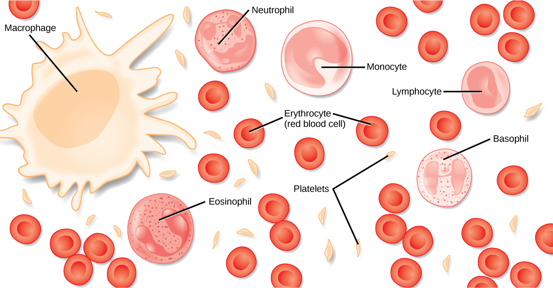
Claret is considered a connective tissue because it has a matrix, every bit shown in (Figure). The living jail cell types are cerise blood cells (RBC), also called erythrocytes, and white claret cells (WBC), likewise chosen leukocytes. The fluid portion of whole blood, its matrix, is normally chosen plasma.
Blood is a connective tissue that has a fluid matrix, called plasma, and no fibers. Erythrocytes (red blood cells), the predominant cell type, are involved in the transport of oxygen and carbon dioxide. Also present are various leukocytes (white blood cells) involved in immune response.
The cell found in greatest abundance in blood is the erythrocyte. Erythrocytes are counted in millions in a blood sample: the boilerplate number of cherry blood cells in primates is iv.7 to v.5 one thousand thousand cells per microliter. Erythrocytes are consistently the same size in a species, but vary in size between species. For example, the boilerplate diameter of a primate cherry-red claret prison cell is 7.five µl, a dog is close at 7.0 µl, but a cat'south RBC diameter is five.9 µl. Sheep erythrocytes are even smaller at iv.6 µl. Mammalian erythrocytes lose their nuclei and mitochondria when they are released from the bone marrow where they are made. Fish, amphibian, and avian ruddy blood cells maintain their nuclei and mitochondria throughout the cell's life. The principal job of an erythrocyte is to carry and evangelize oxygen to the tissues.
Leukocytes are the predominant white claret cells found in the peripheral blood. Leukocytes are counted in the thousands in the blood with measurements expressed as ranges: primate counts range from four,800 to 10,800 cells per µl, dogs from 5,600 to 19,200 cells per µl, cats from 8,000 to 25,000 cells per µl, cattle from four,000 to 12,000 cells per µl, and pigs from 11,000 to 22,000 cells per µl.
Lymphocytes role primarily in the immune response to foreign antigens or material. Dissimilar types of lymphocytes make antibodies tailored to the foreign antigens and control the production of those antibodies. Neutrophils are phagocytic cells and they participate in one of the early lines of defense against microbial invaders, aiding in the removal of bacteria that has entered the body. Some other leukocyte that is found in the peripheral blood is the monocyte. Monocytes give rise to phagocytic macrophages that clean upwardly expressionless and damaged cells in the body, whether they are foreign or from the host animal. 2 additional leukocytes in the blood are eosinophils and basophils—both help to facilitate the inflammatory response.
The slightly granular material among the cells is a cytoplasmic fragment of a prison cell in the bone marrow. This is called a platelet or thrombocyte. Platelets participate in the stages leading upwards to coagulation of the blood to stop haemorrhage through damaged claret vessels. Claret has a number of functions, but primarily it transports material through the body to bring nutrients to cells and remove waste material material from them.
Muscle Tissues
There are three types of muscle in fauna bodies: smooth, skeletal, and cardiac. They differ by the presence or absence of striations or bands, the number and location of nuclei, whether they are voluntarily or involuntarily controlled, and their location within the body. (Figure) summarizes these differences.
| Types of Muscles | ||||
|---|---|---|---|---|
| Blazon of Muscle | Striations | Nuclei | Control | Location |
| smoothen | no | single, in center | involuntary | visceral organs |
| skeletal | yes | many, at periphery | voluntary | skeletal muscles |
| cardiac | yes | unmarried, in center | involuntary | centre |
Smooth Muscle
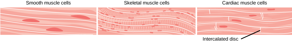
Smooth muscle does not have striations in its cells. Information technology has a single, centrally located nucleus, as shown in (Figure). Constriction of polish muscle occurs under involuntary, autonomic nervous command and in response to local conditions in the tissues. Smoothen muscle tissue is too called non-striated as it lacks the banded advent of skeletal and cardiac muscle. The walls of blood vessels, the tubes of the digestive system, and the tubes of the reproductive systems are composed of by and large smooth muscle.
Smooth muscle cells do not accept striations, while skeletal muscle cells do. Cardiac musculus cells accept striations, simply, unlike the multinucleate skeletal cells, they have only one nucleus. Cardiac muscle tissue also has intercalated discs, specialized regions running along the plasma membrane that bring together adjacent cardiac muscle cells and assist in passing an electric impulse from cell to jail cell.
Skeletal Muscle
Skeletal musculus has striations across its cells caused by the arrangement of the contractile proteins actin and myosin. These muscle cells are relatively long and accept multiple nuclei along the edge of the jail cell. Skeletal muscle is nether voluntary, somatic nervous system control and is found in the muscles that move bones. (Effigy) illustrates the histology of skeletal muscle.
Cardiac Muscle
Cardiac muscle, shown in (Figure), is plant only in the heart. Like skeletal musculus, it has cross striations in its cells, simply cardiac muscle has a single, centrally located nucleus. Cardiac muscle is non under voluntary control merely tin can be influenced by the autonomic nervous system to speed up or slow downwardly. An added characteristic to cardiac musculus cells is a line than extends along the end of the cell as information technology abuts the next cardiac cell in the row. This line is chosen an intercalated disc: it assists in passing electrical impulse efficiently from one prison cell to the next and maintains the strong connectedness between neighboring cardiac cells.
Nervous Tissues
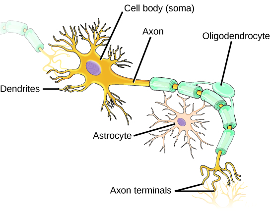
Nervous tissues are fabricated of cells specialized to receive and transmit electric impulses from specific areas of the body and to send them to specific locations in the torso. The master jail cell of the nervous system is the neuron, illustrated in (Figure). The large construction with a central nucleus is the prison cell trunk of the neuron. Projections from the cell body are either dendrites specialized in receiving input or a single axon specialized in transmitting impulses. Some glial cells are also shown. Astrocytes regulate the chemical environment of the nerve jail cell, and oligodendrocytes insulate the axon so the electrical nerve impulse is transferred more efficiently. Other glial cells that are not shown support the nutritional and waste requirements of the neuron. Some of the glial cells are phagocytic and remove debris or damaged cells from the tissue. A nerve consists of neurons and glial cells.
The neuron has projections called dendrites that receive signals and projections chosen axons that send signals. Also shown are two types of glial cells: astrocytes regulate the chemic environment of the nervus cell, and oligodendrocytes insulate the axon so the electrical nervus impulse is transferred more than efficiently.
Link to Learning
Click through the interactive review to acquire more about epithelial tissues.
Career Connections
PathologistA pathologist is a medical doctor or veterinarian who has specialized in the laboratory detection of affliction in animals, including humans. These professionals complete medical school education and follow it with an extensive post-graduate residency at a medical center. A pathologist may oversee clinical laboratories for the evaluation of torso tissue and claret samples for the detection of affliction or infection. They examine tissue specimens through a microscope to identify cancers and other diseases. Some pathologists perform autopsies to determine the cause of death and the progression of disease.
Section Summary
The basic building blocks of complex animals are 4 principal tissues. These are combined to course organs, which accept a specific, specialized function within the body, such equally the skin or kidney. Organs are organized together to perform common functions in the form of systems. The 4 primary tissues are epithelia, connective tissues, muscle tissues, and nervous tissues.
Visual Connexion Questions
(Figure) Which of the following statements most types of epithelial cells is false?
- Elementary columnar epithelial cells line the tissue of the lung.
- Simple cuboidal epithelial cells are involved in the filtering of blood in the kidney.
- Pseudostratisfied columnar epithilia occur in a single layer, just the arrangement of nuclei makes it appear that more than one layer is present.
- Transitional epithelia modify in thickness depending on how full the bladder is.
(Figure) A
Review Questions
Which blazon of epithelial cell is all-time adapted to aid diffusion?
- squamous
- cuboidal
- columnar
- transitional
C
Which blazon of epithelial prison cell is found in glands?
- squamous
- cuboidal
- columnar
- transitional
B
Which blazon of epithelial cell is found in the urinary bladder?
- squamous
- cuboidal
- columnar
- transitional
D
Which blazon of connective tissue has the most fibers?
- loose connective tissue
- fibrous connective tissue
- cartilage
- bone
B
Which type of connective tissue has a mineralized unlike matrix?
- loose connective tissue
- fibrous connective tissue
- cartilage
- os
D
The cell found in bone that breaks it downwardly is called an ________.
- osteoblast
- osteocyte
- osteoclast
- osteon
C
The prison cell found in bone that makes the bone is called an ________.
- osteoblast
- osteocyte
- osteoclast
- osteon
A
Plasma is the ________.
- fibers in claret
- matrix of blood
- jail cell that phagocytizes bacteria
- prison cell fragment found in the tissue
B
The type of muscle cell under voluntary control is the ________.
- smooth muscle
- skeletal muscle
- cardiac musculus
- visceral musculus
B
The part of a neuron that contains the nucleus is the
- jail cell body
- dendrite
- axon
- glial
A
Why are intercalated discs essential to the function of cardiac muscle?
- The discs maintain the barriers between the cells.
- The discs pass nutrients between cells.
- The discs ensure that all the cardiac muscle cells beat as a single unit.
- The discs control the heart rate.
C
Critical Thinking Questions
How can squamous epithelia both facilitate diffusion and prevent damage from abrasion?
Squamous epithelia can be either uncomplicated or stratified. Every bit a single layer of cells, it presents a very sparse epithelia that minimally inhibits diffusion. As a stratified epithelia, the surface cells can be sloughed off and the cells in deeper layers protect the underlying tissues from damage.
What are the similarities betwixt cartilage and os?
Both contain cells other than the traditional fibroblast. Both have cells that lodge in spaces within the tissue called lacunae. Both collagen and elastic fibers are found in os and cartilage. Both tissues participate in vertebrate skeletal development and formation.
Multiple sclerosis is a debilitating autoimmune illness that results in the loss of the insulation around neuron axons. What cell type is the immune system attacking, and how does this disrupt the transfer of messages past the nervous organisation?
In multiple sclerosis, the immune system attacks the oligodendrocytes. The expiry of oligodendrocytes results in the loss of the insulating sheath around the axon of the neurons. When the sheath is gone, the electric impulses travel much more than slowly down the length of the axon.
When a person leads a sedentary life his skeletal muscles atrophy, but his shine muscles practice not. Why?
Skeletal muscles are involved in voluntary move, then the person has to make the pick to work those muscles through do or movement. Smooth muscles are involved in involuntary activities of the body (ex. claret vessel expansion and contraction, abdominal peristalsis) so they are active fifty-fifty when a person is sedentary.
Glossary
- canaliculus
- microchannel that connects the lacunae and aids diffusion betwixt cells
- cartilage
- type of connective tissue with a large corporeality of ground substance matrix, cells called chondrocytes, and some amount of fibers
- chondrocyte
- cell institute in cartilage
- columnar epithelia
- epithelia made of cells taller than they are wide, specialized in absorption
- connective tissue
- type of tissue made of cells, ground substance matrix, and fibers
- cuboidal epithelia
- epithelia made of cube-shaped cells, specialized in glandular functions
- epithelial tissue
- tissue that either lines or covers organs or other tissues
- fibrous connective tissue
- type of connective tissue with a high concentration of fibers
- lacuna
- space in cartilage and bone that contains living cells
- loose (areolar) connective tissue
- type of connective tissue with small amounts of cells, matrix, and fibers; establish effectually blood vessels
- matrix
- component of connective tissue made of both living and nonliving (basis substances) cells
- osteon
- subunit of meaty bone
- pseudostratified
- layer of epithelia that appears multilayered, but is a simple covering
- unproblematic epithelia
- single layer of epithelial cells
- squamous epithelia
- type of epithelia made of flat cells, specialized in aiding diffusion or preventing abrasion
- stratified epithelia
- multiple layers of epithelial cells
- trabecula
- tiny plate that makes up spongy os and gives it strength
- transitional epithelia
- epithelia that can transition for actualization multilayered to simple; besides called uroepithelial
Source: https://opentextbc.ca/biology2eopenstax/chapter/animal-primary-tissues/
Posted by: dudleywainvis1959.blogspot.com
0 Response to "What Are The Types Of Tissues In Animals"
Post a Comment